
Gallo Lab
The Gallo Laboratory investigates postnatal neural development and how injury or disease affects the growth, regeneration and function of neurons and glial cells. Our multidisciplinary research explores the cellular and molecular mechanisms that shape brain development under both normal and pathological conditions.
We are particularly interested in the signals and pathways that regulate the development of neurons and glia within the white matter, cortex and cerebellum, and in translating this knowledge into strategies for cell repair and regeneration following brain injury.
To achieve these goals, we employ an integrated set of molecular, cellular, anatomical, electrophysiological and behavioral approaches, using animal models that replicate key aspects of human brain injury and disease — including perinatal brain injury (such as hypoxia, maternal immune activation, and oxidative stress), Down syndrome and multiple sclerosis.
A major focus of our work is understanding the response of neural progenitor cells to injury and disease and identifying novel molecular mechanisms that promote neural repair and functional recovery.
More recently, our studies have expanded to combine genetically tractable animal models — which enable precise manipulation of signaling pathways — with investigations in large mammals, whose brain anatomy and cellular organization closely resemble those of humans. We have established preclinical models of perinatal brain injury that mimic the gray and white matter alterations observed in very low birth weight (VLBW) premature infants.
Current projects aim to uncover the cellular and physiological bases of cognitive, behavioral and motor deficits in premature infants, with a special focus on the cerebellum and its roles in brain function. Our ongoing studies integrate findings from animal models and postmortem human brain analyses to advance our understanding of neural development, injury and repair.
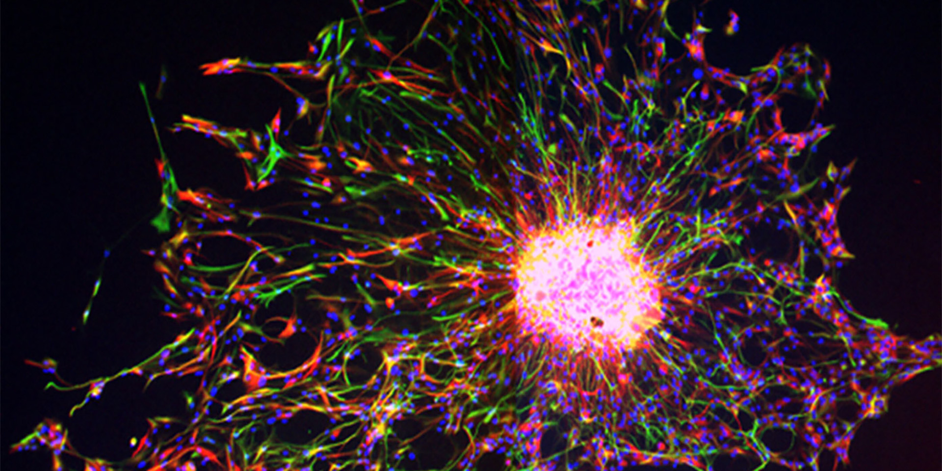
The image above is of the dorsal subventricular zone in an animal model (postnatal day 18) showing expression of Endothelin-1 (green), Glial fibrillary acidic protein (GFAP, red) and cell nuclei (DAPI, blue). GFAP+ astrocytes express the Endothelin-1 protein.
Panagiotis Kratimenos, MD, PhD
Center for Neuroscience Research
Children’s National Research Institute
Division of Neonatology
Children’s National Hospital
Ioannis Koutroulis, MD, PhD, MBA
Center for Genetic Medicine Research
Children’s National Research Institute
Division of Emergency Medicine
Children’s National Hospital
Nobuyuki Ishibashi, MD
Center for Neuroscience Research
Children’s National Research Institute
Children’s National Hospital
Tarik Haydar, PhD
Center for Neuroscience Research
Children’s National Research Institute
Children’s National Hospital
Jeff Dupree, PhD
Department of Anatomy and Neurobiology
Virginia Commonwealth University
Developmental Myelination and Myelin Regeneration